

























Somewhere between the last regular period and the years that follow it, the skeleton quietly changes course. Bone that was once replaced almost as fast as it broke down starts to fall behind, and the gap widens fastest in the first several years after menopause. Estrogen is the hormone most responsible for keeping that balance in check, and when its levels fall, the cells that break down bone gain a temporary advantage over the cells that build it back up.
This article looks at the mechanism behind that shift, why heavy, progressive strength training has become one of the more studied non-drug tools for slowing it, and how a program built around axial loading (loading through the spine and hips, where fracture risk matters most) might be structured. It also covers bone density screening with DEXA and the questions worth bringing to a physician before starting a heavier lifting program. It is written for perimenopausal and postmenopausal women broadly, whether or not a diagnosis of osteopenia or osteoporosis has been made, and it is not a substitute for individualized medical advice.
How Estrogen Decline Changes Bone Remodeling
Bone is not a static scaffold. Throughout adult life, specialized cells called osteoclasts dissolve small amounts of old bone tissue while osteoblasts fill the resulting cavities with new bone. In premenopausal women, estrogen keeps osteoclast activity in check, largely by limiting a signaling molecule called RANKL that recruits and activates these bone-resorbing cells. The result, under normal conditions, is a remodeling cycle where formation roughly keeps pace with resorption.
When ovarian estrogen production drops during the menopause transition, that brake weakens. Osteoclasts become more numerous and more active, resorption outpaces formation, and the skeleton loses density at a rate that is unlike anything seen earlier in adult life. According to the Endocrine Society’s patient education materials, roughly one in two postmenopausal women will go on to develop osteoporosis over their lifetime, a reflection of how significant this shift can be. Cleveland Clinic notes that some women lose up to 20% of their bone density within the first five to seven years following the final menstrual period, with the most rapid losses concentrated in the earliest postmenopausal years before the rate of loss slows to something closer to the gradual age-related decline seen in men.
The bone lost during this window is not distributed evenly. Trabecular bone, the honeycomb-like tissue inside the vertebrae and at the ends of long bones, has a larger surface area relative to its volume and turns over faster than the denser cortical bone that forms the shafts of long bones. That is part of why the spine and hip are watched so closely during this life stage: they contain a higher proportion of trabecular bone and are also the sites where fractures carry the most serious consequences for mobility and independence.
Why This Window Matters
Peak bone mass is typically reached by the late twenties or early thirties. From that point, bone density holds fairly steady until the menopause transition accelerates the loss. Because the years immediately following the final period concentrate so much of a woman’s lifetime bone loss into a short window, what happens during and shortly after that transition can matter more for long-term skeletal health than a decade of ordinary aging on either side of it. This is also the period when many women first hear about bone density testing, T-scores, and fracture risk in concrete terms rather than abstract ones.
Why Strength Training Is Particularly Protective
Bone tissue responds to mechanical strain through a process researchers describe with Frost’s mechanostat theory: bone cells sense the load placed on them and adjust bone mass to better withstand similar loads in the future. Walking and other low-impact activities provide some of this stimulus, but the strain magnitude tends to fall below the threshold needed to meaningfully change bone density in adults, particularly at the hip and spine. Higher-magnitude, progressively increasing loads — the kind produced by resistance training performed close to an individual’s current strength capacity — appear to cross that threshold more reliably.
The clearest evidence for this comes from the LIFTMOR trial, published in the Journal of Bone and Mineral Research in 2018. Researchers screened 568 applicants and enrolled 101 postmenopausal women over age 58 with low bone mass (T-scores below -1.0 at the hip or spine). One group performed twice-weekly, 30-minute supervised sessions of high-intensity resistance and impact training (HiRIT) — five sets of five repetitions at 80 to 85% of one-repetition maximum across deadlifts, overhead presses, and back squats, along with jumping chin-ups for impact loading. The comparison group performed a lower-intensity, home-based program using resistance under 60% of one-repetition maximum. After eight months, the HiRIT group showed roughly 4% greater lumbar spine bone mineral density and about 2% greater femoral neck bone mineral density compared with the control group, along with improvements in functional measures like balance and lower-body power. Notably, the trial reported no serious adverse events in the supervised, carefully screened HiRIT group, though the researchers were explicit that participants had been screened out if they had recent fractures, uncontrolled cardiovascular disease, or other contraindications to heavy loading — a detail that matters for anyone considering a similar program without equivalent screening and supervision.
A more recent systematic review and meta-analysis of 17 randomized controlled trials, covering 690 postmenopausal participants and published in 2025, examined which training variables most consistently improved bone mineral density. Training performed three times per week produced significant improvements at the lumbar spine, femoral neck, total hip, and trochanter. High-intensity protocols at or above 70% of one-repetition maximum showed a significant effect at the total hip and femoral neck specifically, and interventions lasting 48 weeks or longer produced better outcomes at the femoral neck and total hip than shorter programs. The review’s authors concluded that a high-intensity regimen performed three times weekly over a longer duration appeared optimal, while noting meaningful variability between the underlying studies.
Taken together, these findings point toward a pattern: modest loads and casual frequency are unlikely to move the needle much on bone density at the hip and spine, but structured, progressively heavier resistance training performed multiple times a week over many months has produced measurable, site-specific gains in trials of postmenopausal women.
Building a Program: Frequency, Intensity, and Exercise Selection
None of this is a prescription to follow without individualized guidance, particularly for anyone with an existing osteoporosis diagnosis, a prior fragility fracture, or other health conditions that affect exercise tolerance. It does, however, outline the shape that research-informed programming tends to take.
Frequency
Across the trials described above, two to three sessions per week focused on resistance training was the range associated with meaningful bone density change. Fewer than two sessions weekly, or programs using only light resistance, have not shown the same consistent effect on hip and spine density in this population.
Intensity and Progression
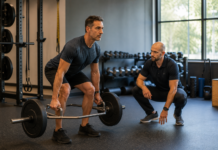
The studies with the clearest bone density benefit used loads in the range of 70 to 85% of one-repetition maximum — heavy enough that the last one or two repetitions of a set are genuinely difficult to complete with good form. This is a meaningfully heavier load than the light resistance bands or one- to three-kilogram weights sometimes marketed toward older women. Reaching that intensity safely is a gradual process: the LIFTMOR protocol itself used a full month of lighter, bodyweight-based preparation before progressing to the heavier working loads, and any similar transition should be built with professional guidance, ideally from a physical therapist or certified trainer experienced in working with older adults or people with low bone density.
Exercise Selection: Favoring Axial Loading
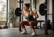
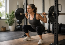
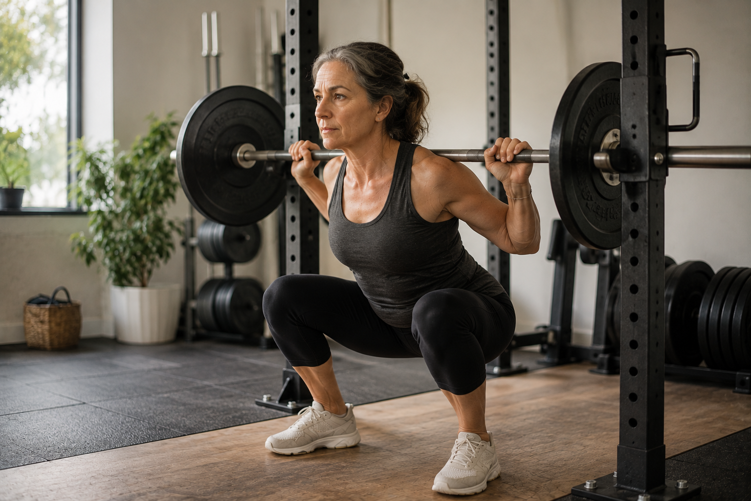
Because trabecular bone loss concentrates in the spine and hip, exercises that load the body along its vertical axis tend to be emphasized in bone-focused programs. Barbell or dumbbell back squats, deadlifts (including trap-bar variations, which can reduce forward spinal flexion), and overhead presses direct compressive force through the spine and hip joints in a controlled, upright position. Impact-based movements such as low-level jumping or hopping, when appropriate for the individual’s fracture risk and cleared by a physician, add a further loading stimulus that walking alone does not provide.
The Bone Health & Osteoporosis Foundation draws an important distinction here: not every form of movement traditionally recommended for older adults is automatically bone-protective, and some popular flexibility practices carry their own cautions. The Foundation specifically notes that certain yoga and Pilates positions involving forward spinal flexion or twisting may increase the risk of vertebral fracture in people with low bone density, and recommends working with a physical therapist to identify safe variations rather than assuming all “gentle” exercise is equally protective of the spine.
Program Guidance Snapshot
- Frequency: 2-3 resistance sessions per week, non-consecutive days
- Intensity: Progress toward 70-85% of one-repetition maximum, working with a trainer or physical therapist to build up safely
- Core movements: Squat, deadlift or trap-bar deadlift, overhead press, row — patterns that load the spine and hip axially
- Duration: Sustained programs of 8-12+ months, since bone remodeling occurs on a slower timescale than muscle adaptation
- Caution: Avoid unsupervised heavy spinal flexion or twisting loads if bone density is already low or a fracture history exists
- Before starting: Confirm with a physician, especially with a prior fracture, cardiovascular condition, or existing osteoporosis diagnosis
Muscle, Balance, and Fall Prevention
Bone density is only part of the fracture equation. Most fractures in older women happen as the result of a fall, not from bone failing on its own during normal daily movement, which means the muscle strength and balance built during resistance training carry protective value beyond what shows up on a DEXA scan. Sarcopenia, the age-related loss of muscle mass and strength, tends to accelerate around the same postmenopausal window as bone loss, and the two processes appear to reinforce each other: weaker muscles produce less mechanical pull on bone, and lower bone density can make a person more cautious and less physically active, which further reduces the loading stimulus muscles and bone both need.
Compound lifts such as squats and deadlifts train the muscle groups most responsible for catching a stumble before it becomes a fall, including the hip extensors, quadriceps, and the deep stabilizing muscles around the ankle and knee. Programs that pair resistance training with a balance component, such as single-leg stance work or controlled step-downs, address both sides of the fracture-risk equation at once: the strength to move safely and the bone density to withstand an impact if a fall does happen. This dual benefit is one reason strength training is frequently recommended alongside, rather than instead of, other components of a bone-health plan.
Supporting Nutrients: Calcium, Vitamin D, and Protein
Strength training builds bone most effectively when the raw materials for bone formation are available, and the Endocrine Society specifically pairs its bone-loss guidance with calcium and vitamin D intake, noting that vitamin D is what allows the body to absorb dietary calcium in the first place. Without adequate vitamin D, additional calcium intake does relatively little to support bone mineralization, regardless of how much resistance training is performed. A physician or registered dietitian can help identify whether blood levels of vitamin D are adequate and whether dietary intake of calcium, through foods like dairy, fortified plant milks, and leafy greens, is meeting individual needs, since requirements vary by age, existing bone density, and other health conditions.
Adequate dietary protein also supports the muscle-building side of a resistance training program, and postmenopausal women are frequently advised to pay closer attention to protein intake as part of counteracting age-related muscle loss. None of these nutritional factors substitute for the mechanical loading strength training provides, but they form the supporting environment that allows bone and muscle to respond to that loading.
DEXA Screening and Knowing Where You Stand
Dual-energy X-ray absorptiometry, or DEXA (sometimes written DXA), is the standard imaging test used to measure bone mineral density, most commonly at the hip and lumbar spine. Results are reported as a T-score, comparing an individual’s bone density to that of a healthy young adult; a T-score of -1.0 or above is considered normal, between -1.0 and -2.5 indicates osteopenia (low bone mass), and -2.5 or below indicates osteoporosis.
The U.S. Preventive Services Task Force recommends bone density screening for all women age 65 and older, and for postmenopausal women younger than 65 who have risk factors that raise their fracture risk above that of an average 65-year-old — factors such as low body weight, a parent’s hip fracture history, smoking, or certain medications. A DEXA scan taken around the time of menopause, even before age 65, can establish a personal baseline, which makes it easier to see whether bone density is declining faster than expected and whether a given exercise and lifestyle approach is having the intended effect over time.
Working With a Physician
Strength training research in postmenopausal women has consistently been conducted under medical screening and often direct supervision, and that context matters when translating trial results into an individual program. A physician can review fracture risk, order or interpret a DEXA scan, check for conditions like vitamin D deficiency or thyroid disorders that affect bone metabolism, and flag any reason a heavier lifting program might need modification, such as an existing vertebral fracture, uncontrolled hypertension, or a recent joint replacement. Where bone density is already significantly low, some physicians also weigh strength training as a complement to, rather than a replacement for, medications that reduce fracture risk. None of the research described in this article should be read as a reason to delay or avoid a conversation with a physician or bone health specialist, particularly for anyone with a known diagnosis of osteopenia or osteoporosis, a prior fracture, or symptoms like unexplained back pain or height loss.
Conclusion
Bone density decline after menopause follows a predictable biological path: falling estrogen loosens the restraint on cells that break down bone, and the spine and hip lose density faster than they gain it back. Progressive, heavy resistance training is one of the more thoroughly studied ways to push back against that imbalance, with trial evidence pointing toward two to three sessions weekly, loads in the 70 to 85% of one-repetition maximum range, and axial movements like squats, deadlifts, and presses sustained over many months. A DEXA scan provides the baseline and follow-up data to know whether an individual program is working, and a physician remains the right person to confirm that a given plan fits an individual’s fracture risk, medical history, and current bone density.
Frequently Asked Questions
Is strength training alone enough to prevent osteoporosis after menopause?
Strength training is one part of a broader approach that typically also includes adequate calcium and vitamin D intake, fall-prevention strategies, and, for some women, medication. It has not been shown to fully reverse established osteoporosis on its own, and a physician should help determine whether medication is also appropriate based on DEXA results and fracture risk.
What if I have already been diagnosed with osteoporosis: can I still lift heavy weights?
Trials like LIFTMOR specifically included women with osteopenia and osteoporosis, and found the supervised, screened program to be well tolerated. That said, participants were carefully screened for contraindications and worked with qualified supervision. Anyone with an existing osteoporosis diagnosis should have an exercise plan reviewed by a physician or physical therapist before beginning heavier loading.
How soon after menopause should I get a DEXA scan?
Guidelines call for screening at age 65 for average-risk women, and earlier for postmenopausal women with additional risk factors. Many women choose to discuss a baseline scan with their physician around the time of menopause, particularly if risk factors are present, so that future changes can be measured against a known starting point.
Can walking or yoga replace strength training for bone health?
Walking and yoga offer real benefits for cardiovascular health, balance, and flexibility, but the loading forces involved are generally lower than what trials have found necessary to change bone density at the hip and spine. The Bone Health & Osteoporosis Foundation also cautions that some yoga and Pilates positions involving spinal flexion may not be appropriate for people with low bone density.
How long before I would see a change in bone density from strength training?
Bone remodels more slowly than muscle adapts. Trials showing measurable bone mineral density changes generally ran eight months to a year or longer, and the 2025 meta-analysis found that programs of 48 weeks or more produced better hip and femoral neck outcomes than shorter ones.
Do I need a trainer, or can I start this on my own?
Given that the strongest evidence comes from supervised programs with careful screening and technique coaching, working with a physical therapist or a trainer experienced in bone health, at least initially, is a reasonable way to build toward heavier loads safely.
References
- Watson, S.L., et al. “High-Intensity Resistance and Impact Training Improves Bone Mineral Density and Physical Function in Postmenopausal Women With Osteopenia and Osteoporosis: The LIFTMOR Randomized Controlled Trial.” Journal of Bone and Mineral Research, 2018. onlinelibrary.wiley.com/doi/abs/10.1002/jbmr.3284
- “Optimal resistance training parameters for improving bone mineral density in postmenopausal women: a systematic review and meta-analysis.” PMC, National Library of Medicine. pmc.ncbi.nlm.nih.gov/articles/PMC12107943
- Bone Health & Osteoporosis Foundation. “Be Bone Strong: Weight-Bearing and Muscle-Strengthening Exercises.” bonehealthandosteoporosis.org
- Endocrine Society. “Menopause and Bone Loss.” Endocrine Library, Patient Engagement. endocrine.org/patient-engagement/endocrine-library/menopause-and-bone-loss
- Cleveland Clinic. “What To Know About Menopause and Bone Loss.” health.clevelandclinic.org/osteoporosis-and-menopause
- U.S. Preventive Services Task Force. “Screening for Osteoporosis to Prevent Fractures: Recommendation Statement.” uspreventiveservicestaskforce.org/uspstf/recommendation/osteoporosis-screening
- The Menopause Society (North American Menopause Society). Position Statements Collection. Menopause journal. journals.lww.com/menopausejournal/collections/1